Glycolipids as key molecules
Since a large number of viral and bacterial zoonotic agents and the virulence factors (toxins) they produce bind to glycoconjugates on the cell surface of their target cells, this course focused on glycolipids as potential receptor molecules. These consist of a double-tailed lipid anchor, the so-called ceramide (sphingosine plus fatty acid), and an oligosaccharide part, which is characterized by a high variability due to the different number and linkage type of sugars. Glycolipids are found in the outer layer of the plasma membrane of human and animal cells. Consequently, glycolipids play an important role as receptors for zoonotic pathogens and their virulence factors.
The workshop was divided into 2 sections: teaching the theoretical basics on the first day and conducting mass spectrometric experiments in small groups on the second day.
Teaching the theoretical basics

The basic theoretical knowledge on mass spectrometry required for this course was imparted in the form of 8 lectures. First, the structures and receptor functions of glycolipids, especially under zoonotic aspects, were presented in a review lecture (Johannes Müthing). Furthermore, the basic analytical techniques for the subsequent mass spectrometric experiments were explained, whereby the use of thin layer chromatography (DC) coupled with immunochemical detection techniques was described in detail. This was followed by two lectures in which the basics of matrix-assisted laser desorption/ionization mass spectrometry (MALDI-MS) (Klaus Dreisewerd) and electrospray ionization mass spectrometry (ESI-MS) (Michael Mormann) were taught. The lectures on the two fundamental laser and electrospray ionization processes were rounded off with a presentation of the basic principles of mass analysis and the general functioning of mass analyzers (Michael Mormann). Subsequently, selected applications of ESI-MS were presented in two lectures, whereby the mass spectrometric characterization of Shiga toxin receptors (Iris Meisen) and influenza virus receptors (Gottfried Pohlentz) by means of tandem-MS was explained as an example.
MALDI-MS with great potential in zoonoses research
The lipid topic was concluded with a lecture on various applications of MALDI-MS, in which the extraordinary potential of mass spectrometry for structure elucidation not only of glycolipids but also of bioactive lipids of various kinds was demonstrated (Klaus Dreisewerd). The presentation of MALDI-MS as a "new" method for species identification of bacteria impressively illustrated the rapid development of MS-based techniques as the future technologies for numerous applications (Alexander Mellmann). MALDI-MS methods have already largely replaced the "classical" typing methods of pathogens and have found their way into routine microbiological diagnostics. Mass spectrometric technologies will therefore also gain in importance in the future for the identification of zoonotic pathogens.
Literature:
Distler U, Hülsewig M, Souady J, Dreisewerd K, Haier J, Senninger N, Friedrich AW, Karch H, Hillenkamp F, Berkenkamp S, Peter-Katalinić J and Müthing J (2008) Matching IR-MALDI-o-TOF mass spectrometry with the TLC overlay binding assay and its clinical application for tracing tumor-associated glycosphingolipids in hepatocellular and pancreatic cancer. Anal Chem 80, 1835-1846.
Dreisewerd K, Müthing J, Rohlfing A, Meisen I, Vukelić Ž, Peter-Katalinić J, Hillenkamp F and Berkenkamp S (2005) Analysis of gangliosides directly from thin-layer chromatography plates by infrared matrix-assisted laser desorption/ionization orthogonal-time-of-flight mass spectrometry. Anal Chem 77, 4098-4107.
Meisen I, Dzudzek T, Ehrhardt C, Ludwig S, Mormann M, Lümen R, Kniep B, Karch H and Müthing J (2012) The human H3N2 influenza A viruses A/Victoria/3/75 and A/Hiroshima/52/2005 preferentially bind to α2,3-sialylated monosialogangliosides with fucosylated poly-N-acetyllactosaminyl chains. Glycobiology 22, 1055-1076.
Meisen I, Mormann M and Müthing J (2011) Thin-layer chromatography, overlay technique and mass spectrometry: a versatile triad advancing glycosphingolipidomics. Biochim Biophys Acta 1811, 875-896.
Mellmann A and Müthing J. MALDI-TOF mass spectrometry-based microbial identification. In: Advanced Techniques in Diagnostic Microbiology, YW Tang and CW Stratto (eds.), Springer Science+Business Media New York 2012, DOI 10.1007/978-1-4614-3970-7_10.
Müsken A, Souady J, Dreisewerd K, Zhang W, Distler U, Peter-Katalinić J, Miller-Podraza H, Karch H and Müthing J (2010) Application of thin-layer chromatography/infrared matrix-assisted laser desorption/ionization orthogonal time-of-flight mass spectrometry to structural analysis of bacteria-binding glycosphingolipids selected by affinity detection. Rapid Commun Mass Spectrom 24, 1032-1038.
Müthing J and Distler U (2010) Advances on the compositional analysis of glycosphingolipids combining thin-layer chromatography with mass spectrometry. Mass Spectrom Rev 29, 425-479.
Müthing J, Meisen I, Zhang W, Bielaszewska M, Mormann M, Bauerfeind R, Schmidt MA, Friedrich AW and Karch H (2012). Promiscuous Shiga toxin 2e and its intimate relationship to Forssman. Glycobiology 22, 849-862.
Putting the acquired knowledge into practice
The participants first carried out preparatory work in small groups for the subsequent MS studies of glycolipids. A reference glycolipid mixture of human erythrocytes, which contained the receptors for the Shiga toxins produced by enterohaemorrhagic E. coli (EHEC), was separated by thin layer chromatography (DC). The glycolipids globotriaosylceramide (Gb3Cer) and globotetraosylceramide (Gb4Cer), which are the receptors for Shiga toxins, were detected immunochemically on the DC plate. Subsequently, DC strips were prepared for mass spectrometric receptor characterization using MALDI and ESI-MS and the spectra obtained were evaluated.

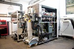
After separation of the glycolipid mixture, the individual glycolipids were first detected with the sugar-specific reagent orcinol (control experiment). The DC overlay assay, which corresponds to the principle of the ELISA test procedure, served as the immunological detection method. Shiga toxin receptors were detected by means of specific anti-Gb3Cer and anti-Gb4Cer antibodies. Antibody binding was detected with the substrate 5-bromo-4-chloro-3'-indolyphosphate. For subsequent direct coupling of the DC overlay assay with MALDI-MS, the DC plate was cut to target size with a glass cutter, while for indirect coupling with ESI-MS the silica gel of the immune-stained glycolipids was scraped off and the glycolipids were subsequently extracted from the silica gel with organic solvents. The immunopositive glycolipid bands of the DC-strip, which was cut to fit the MALDI target, were wetted with glycerol (matrix). The pretreated strip was then introduced into the MALDI mass spectrometer, which was a modified prototype of an orthogonally extracting mass spectrometer equipped with a time-of-flight (ToF) analyzer. In the ion source, the glycolipid band was bombarded with nanosecond laser pulses at a wavelength of 2.94 µm. During the explosive material removal, the glycolipid molecules in the band were desorbed and partially ionized. Based on the different flight times of the formed ions, the mass-to-charge ratios (m/z) of the ions were determined in the time-of-flight analyzer. Since the glycolipids remain intact during the "gentle" ionization by MALDI, the m/z measurement values were used to make structural suggestions for the detected glycolipid species. This included, for example, the determination of the fatty acid (C16 to C24) and the determination of the oligosaccharide type (trisaccharide in the case of Gb3Cer and tetrasaccharide in the case of Gb4Cer). In the course of the workshop the participants were able to operate the device themselves and record mass spectra after a short introduction. These were then evaluated using examples.

In a second experiment, the glycolipids extracted from the silica gel were analysed in more detail by means of ESI-MS. For this purpose the extracts were taken up in methanol with 1% formic acid and the dissolved glycolipids were adjusted to a concentration of 20 pmol/µl. The participants first recorded overview spectra analogous to the MALDI measurements and then assigned the signals to the ionized molecules of the detected glycolipid species. To perform the tandem MS fragmentation experiment by means of "collision-induced dissociation" (CID), which allows an exact structure elucidation, precursor ions with a m/z value of 1361.92 were selected as an example and the corresponding structure of Gb4Cer (d18:1/C24:0) was determined. The variability of the fragmentation efficiency, which is important for a clear structure determination, could be demonstrated by changing the collision energy. Finally, the supervisors explained the fragmentation signals and explained the derivation of the exact structures from the detected B, Y and W'' fragments.
Concluding remarks and outlook
The workshop concept, consisting of theory (lectures) and practice (experiments in small groups), was well received by the participants and the commitment of all involved supervisors was appreciated. During the two-day workshop, lively discussions developed between the participants and the organisers; first cooperations with the MS-Group in Münster were initiated. The inclusion of further members of the National Research Platform for Zoonoses in a future cross-sectional project was intensively discussed in an evening round of talks over traditional Westphalian cuisine.
The workshop was documented photographically by Ivan Kouzel, to whom we would like to take this opportunity to thank him for the photographs.


